Radiology Outpatient Service Hours
7am to 5pm Monday through Friday.
Radiology Office Numbers 918-485-1344 or 918-485-1345.
On Staff Radiologist, Dr. Heath A. Vandelinder, M.D.s
Types of Radiology Exams offered at W.C.H.
MRI, CT, X-ray, Nuclear Medicine, Digital Mammography, Dexa Bone Density
Special Procedures offered in W.C.H. Radiology
Guided Biopsies, Liver, Kidney, and Thyroid. Abcess drainage, Cyst Drainage, Thoracentesis, Paracentesis

Primary Specialty
Diagnostic & Interventional Radiology
Medical School
Touro University College of Osteopathic Medicine, Vallejo, CA
Residency
Oklahoma State University College of Osteopathic Medicine, Tulsa, OK
Fellowship
Advanced Body Imaging Fellowship, University of Southern California
Board Certified
American Osteopathic Board of Radiology
More
Member of Board of Directors, Tulsa Diagnostic & Interventional Radiology

Primary Specialty
Diagnostic & Interventional Radiology
Medical School
Oklahoma State University College of Osteopathic Medicine, Tulsa, OK
Residency
Diagnostic Radiology, Oklahoma State University Medical Center, Tulsa, OK
Fellowship
Advanced Body Imaging Fellowship, University of Southern California
Board Certified
American Osteopathic Board of Radiology
More
Member of Board of Directors, Tulsa Diagnostic & Interventional Radiology

Primary Specialty
Diagnostic & Interventional Radiology
Medical School
University of South Florida College of Medicine, Tampa, FL
Residency
Diagnostic Radiology, Integris Baptist Medical Center Oklahoma City, OK
Board Certified
American Board of Radiology
More
Member of Board of Directors, Tulsa Diagnostic & Interventional Radiology
• CT / Computed Tomography
• CT Angiography including Coronary studies
• Low Dose Lung Screening
• General Head/Spine/Abdominal/Orthopedic
• CT Guided Biopsy
• Mobile MRI Services
• Musculoskeletal
• Head & Spine
• Orthopedic Joint
• MRCP
• 3D Digital Mammography
• Osteoporosis Screenings / DEXA scan
Ultrasound
• Echocardiograms
• Vascular
• OB/GYN
• Thyroid Scan & Biopsy
• Breast Mass Scan & Biopsy
• General Abdominal/Gallbladder/Soft tissue/Scrotum
• Carotid/Aorta/Peripheral Arterial Disease Screenings
Nuclear Medicine
• Thyroid Uptake
• Gallbladder/HIDA
• Bone Scan
• MPI Chemical Stress Test
• Pulmonary Perfusion
Diagnostic Xray Imaging
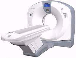
CT Optima 660 64 slice
A comfortable experience The Optima CT660 enables short scan times with 40-mm thin-slice acquisitions for reliable studies. The video of relaxing scenes or cartoons can have a calming effect on children or patients of all ages. An automated voice system provides the ability to give instructions in the patient’s own language. In the low position, the exam table helps with access for patients in wheelchairs.
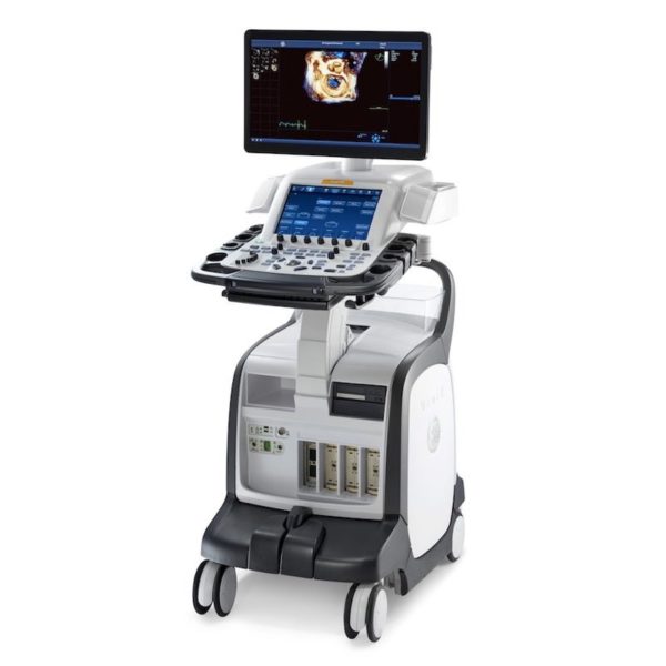
Ultrasound GE Vivid E90
GE Vivid E90 is a premium cardiovascular ultrasound system. The Vivid E90 also covers a wide range of clinical applications through different types of transducers.
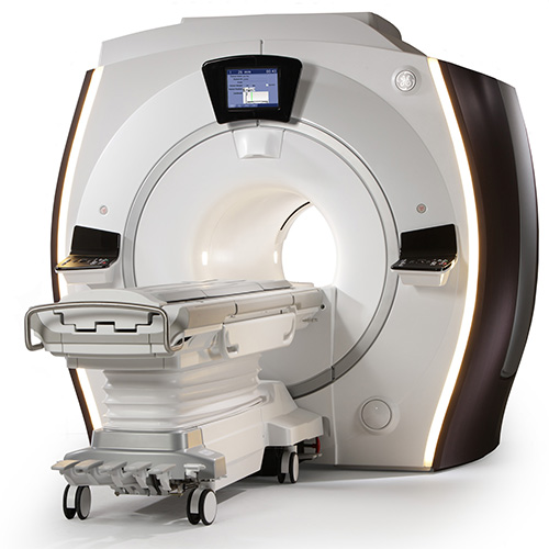
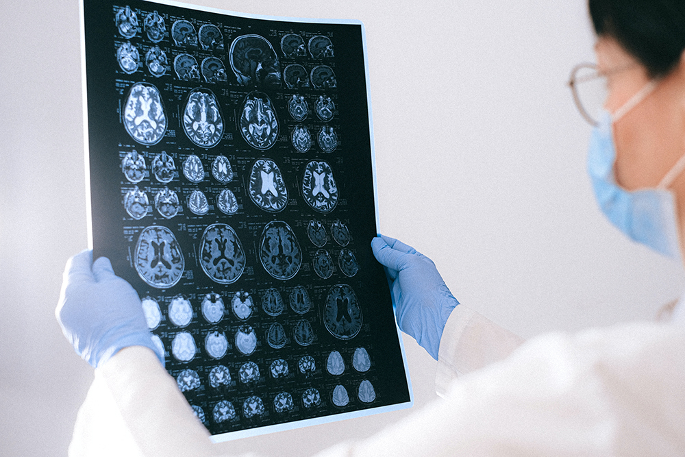
MRI GE 1.5T Magnet
Multi-channel coils
Boosted Speed
High Quality Imaging
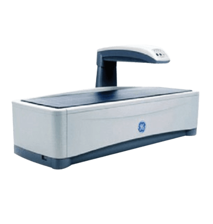
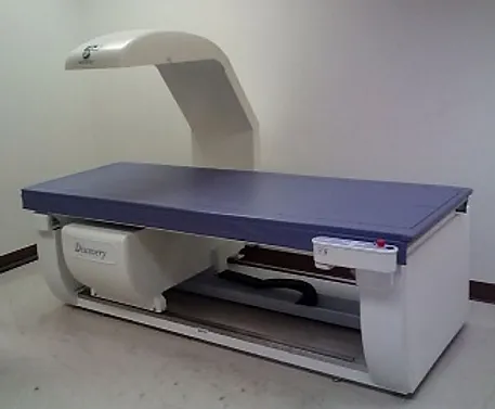
DEXA Prodigy Full Oracle Scanner
Dependable dual-energy X-ray absorptiometry (DXA) assessment, and Prodigy delivers with industry-leading precision and low-dose radiation.
• Total body bone and tissue composition
• Dual-energy vertegral assessment
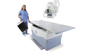
Diagnostic Xray Suite
GE Proteus
High Quality Digital Imaging
Dose Efficient
Nuclear Medicine
GE Millennium MG Dual Head Nuclear Camera with Xeleris 4DR Processing
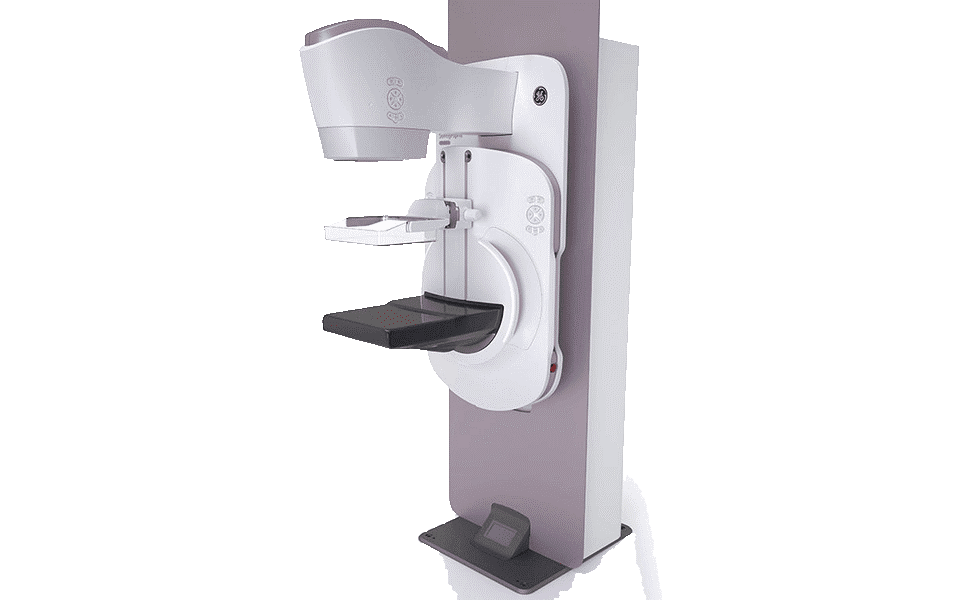
GE Senographe Pristina 3D
Low Dose for Diagnostic Accuracy
Senographe Pristina is the ONLY FDA approved 3D mammography that delivers at the same low dose as 2D FFDM the lowest patient dose of all FDA approved systems

Our bone scans use a technology called DEXA (for dual energy X-ray absorptiometry). In a DEXA scan, you recline on a table while a technician aims a scanner mounted on a long arm.
The DEXA scanner uses beams of very low-energy radiation to determine the density of the bone. The amount of radiation is tiny: about one-tenth of a chest X-ray. The scan is painless, and considered completely safe. Pregnant women should not get DEXA scans because a developing baby shouldn’t be exposed to radiation, no matter how low the dose.
Measurements are usually taken at the hip, and sometimes the spine and other sites. Insurance or Medicare generally pays for the test in women considered at risk for osteoporosis, or those already diagnosed with osteoporosis or osteopenia.
The DEXA can also assess your risk for developing fractures. The risk of fracture is affected by age, body weight, history of prior fracture, family history of osteoporotic fractures and life style issues such as cigarette smoking and excessive alcohol consumption. These factors are taken into consideration when deciding if a patient needs therapy.

Mammograms are used as a screening tool to detect early breast cancer in women experiencing no symptoms. They can also be used to detect and diagnose breast disease in women experiencing symptoms such as a lump, pain, skin dimpling or nipple discharge.
Mammography plays a central part in early detection of breast cancers because it can show changes in the breast up to two years before a patient or physician can feel them. Current guidelines from the U.S. Department of Health and Human Services (HHS) and the American College of Radiology (ACR) recommend screening mammography every year for women, beginning at age 40. Research has shown that annual mammograms lead to early detection of breast cancers, when they are most curable and breast-conservation therapies are available.
The National Cancer Institute (NCI) adds that women who have had breast cancer, and those who are at increased risk due to a family history of breast or ovarian cancer, should seek expert medical advice about whether they should begin screening before age 40 and the need for other types of screening. If you are at high risk for breast cancer, you may need to obtain a breast MRI in addition to your annual mammogram.
